-
-
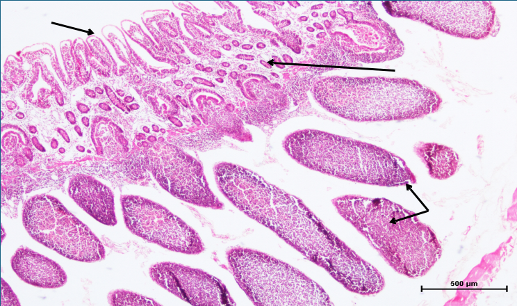
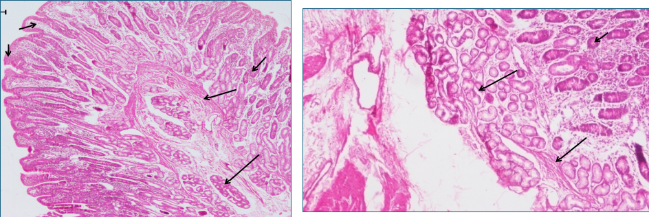
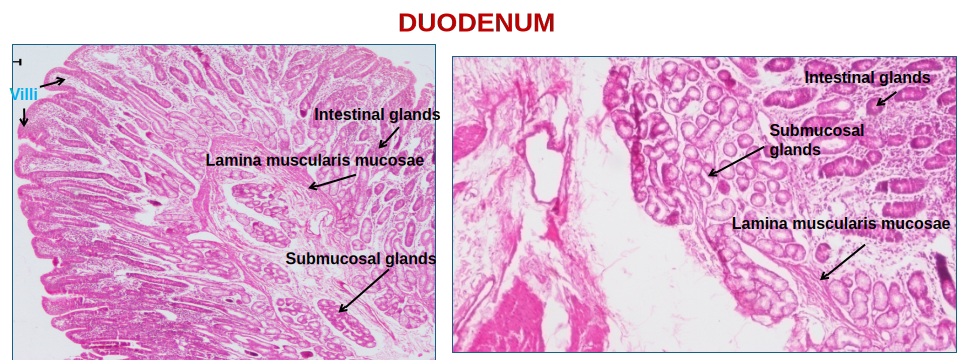
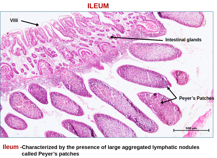 SMALL INTESTINEGeneral Histological organization – Comprised of the duodenum, jejunum, and ileum. The absorptive surface of the small intestine is increased by:•Circular mucosal submucosal folds called plicae circularis•Presence of finger-like mucosal projections called villi and microvilli on the lining epithelial cells.1. Tunica mucosa- Villi (mucosal projections) are the most characteristic feature of the small intestine. The shape of villi is short and wide in ruminants and long and finger-like in carnivoresLamina epithelialis- Simple columnar epithelium having goblet cells in between the lines of villi. The luminal surface of cells is covered with microvilli.Lamina propria- Loose connective tissue that forms the core of villi and surrounds intestinal glands (Crypts of Lieberkuhn). Each villus has a central lymph capillary called a lacteal.Lamina muscularis mucosae- Composed of an inner circular and an outer longitudinal layer of smooth muscle.Subglandular layer between the base of the intestinal glands and the lamina muscularis mucosae in carnivores.2. Tunica submucosa- The initial portion of the small intestine (duodenum) contains submucosal glands (Brunner’s gland), which may continue into the jejunum. Glands are mucous in ruminants and dogs and serous in pigs and horses. Ducts of glands pierce the muscularis mucosae and mostly end up in the intestinal glands. Solitary lymphatic nodules are present throughout the small intestinal submucosa, but large aggregated lymphatic nodules called Peyer’s Patches are numerous in the ileum.3. Tunica muscularis- Inner circular and outer longitudinal layers.4. Tunica serosa- Loose connective tissue covered by mesothelium.