SALIVARY GLANDS
SALIVARY GLANDS: The major salivary glands are parotid, mandibular, and sublingual glands. However, in dog, the zygomatic salivary gland is also present
-
According to the type of secretory cells, salivary glands can be classified into
1.
Serous salivary gland composed of serous secretory cells, e.g., parotid salivary gland.
2.
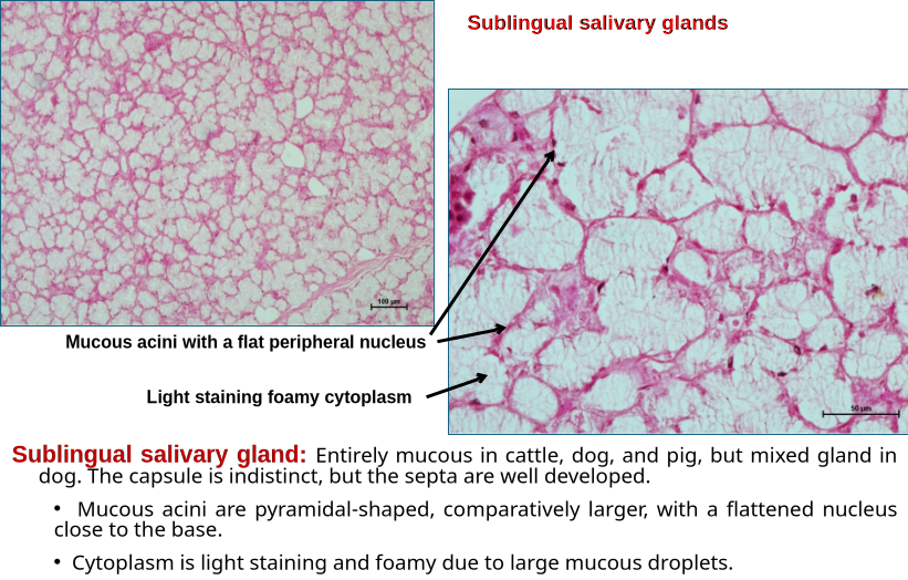
Mucous salivary gland having mucous secretory cells, e.g., the sublingual salivary gland in the cow, sheep, and pig.
3.
Mixed salivary gland having both serous and mucous acini.
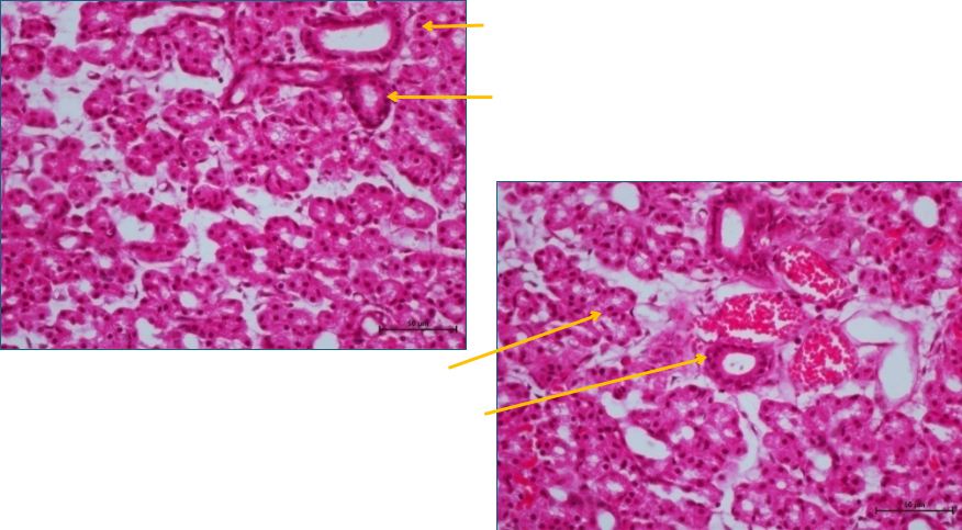
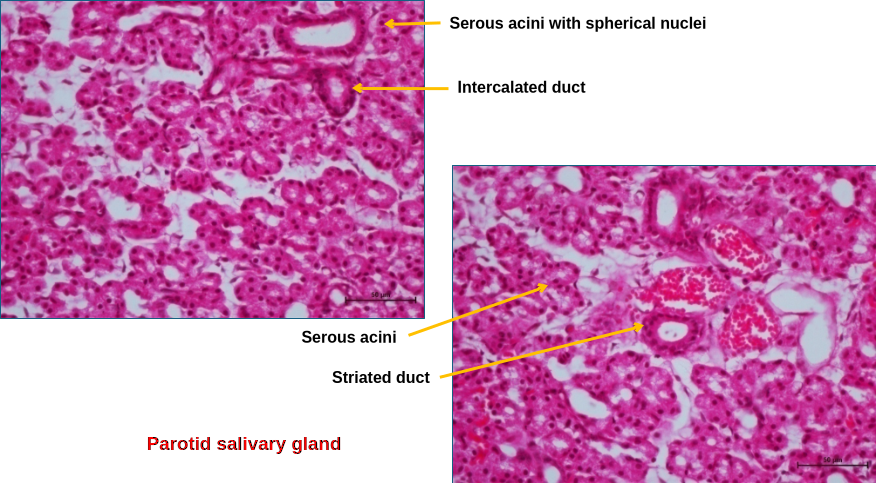
Parotid salivary gland: Surrounded by a connective tissue capsule from which septa radiate into the gland, dividing it into lobes and lobules.
•
Each lobule was composed of serous acini of variable shape and size, ducts, blood vessels, and nerves.
•
Serous acini are lined by pyramidal-shaped cells with spherical nuclei placed close to the base. Apical cytoplasm is filled with zymogen granules. Myoepithelial cells (basket cells) surround the acini, and their contraction facilitates the release and movement of the secretory product
•
The lumen of acini is small and opens into the intercalated duct, which is lined by low cuboidal epithelium.
•
Several of the intercalated ducts come together to form a striated duct, which may be lined by cuboidal or columnar epithelium with striations towards the base.
•
Within lobules, striated ducts merge and form intralobular ducts with stratified cuboidal epithelium, which join with one another to form interlobular ducts having stratified columnar epithelium. Interlobular ducts empty saliva into the interlobar duct, which then empties into the main parotid duct.
 SALIVARY GLANDSSALIVARY GLANDS: The major salivary glands are parotid, mandibular, and sublingual glands. However, in dog, the zygomatic salivary gland is also present-According to the type of secretory cells, salivary glands can be classified into1.Serous salivary gland composed of serous secretory cells, e.g., parotid salivary gland.2.Mucous salivary gland having mucous secretory cells, e.g., the sublingual salivary gland in the cow, sheep, and pig.3.Mixed salivary gland having both serous and mucous acini.Parotid salivary gland: Surrounded by a connective tissue capsule from which septa radiate into the gland, dividing it into lobes and lobules.•Each lobule was composed of serous acini of variable shape and size, ducts, blood vessels, and nerves.•Serous acini are lined by pyramidal-shaped cells with spherical nuclei placed close to the base. Apical cytoplasm is filled with zymogen granules. Myoepithelial cells (basket cells) surround the acini, and their contraction facilitates the release and movement of the secretory product•The lumen of acini is small and opens into the intercalated duct, which is lined by low cuboidal epithelium.•Several of the intercalated ducts come together to form a striated duct, which may be lined by cuboidal or columnar epithelium with striations towards the base.•Within lobules, striated ducts merge and form intralobular ducts with stratified cuboidal epithelium, which join with one another to form interlobular ducts having stratified columnar epithelium. Interlobular ducts empty saliva into the interlobar duct, which then empties into the main parotid duct.
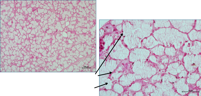
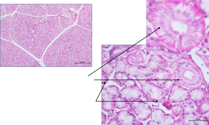
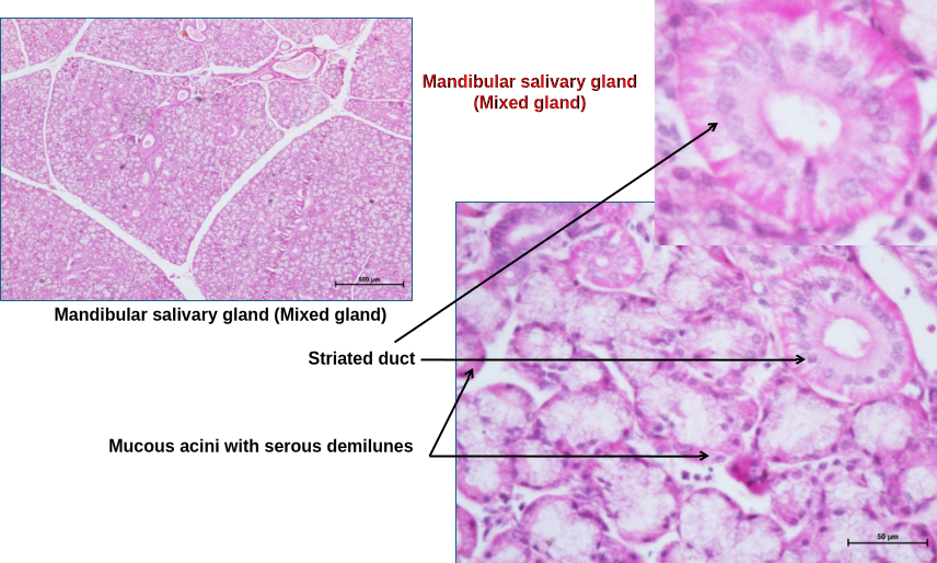 Mandibular salivary glands: Mixed type of gland. The distribution of serous and mucous cells varies across acini.•Some acini are completely serous, and others are purely mucous; however, in some acini, both serous and mucous cells are present.•Some mucous acini have crescent-shaped bodies of serous cells called serous demilunes.