-
-
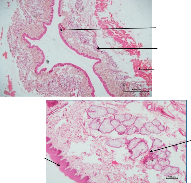
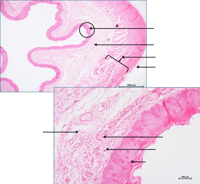
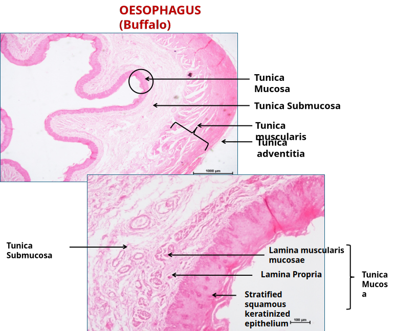
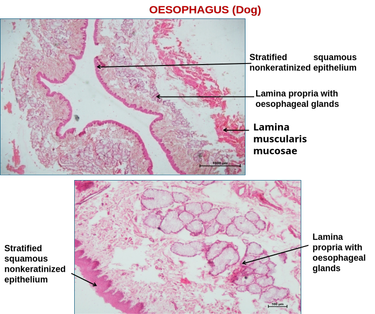 OESOPHAGUSGeneral Histological Organization: Tubular organs of the digestive tract from the esophagus to the rectum are comprised of tunica mucosa, tunica submucosa, tunica muscularis, and tunica serosa or adventitia from the lumen to the outside.1.Tunica mucosa: Further comprises the following three layersi.Lamina epithelialis: It is stratified squamous keratinized in ruminants and non-keratinized in dogs and pigs. The degree of keratinization is related to the texture of the diet.ii.Lamina propria: Comprised of a network of connective tissue fibers.iii.Lamina muscularis mucosae: Comprised of longitudinally oriented smooth muscle bundles. In ruminants and horses, isolated bundles near the pharynx increase towards the stomach. Absent cranially in pigs and dogs.2. Tunica submucosa: Loose connective tissue layer. The glands are present along the entire length of the esophagus in dogs, in the cranial half of the esophagus in pigs, and in the pharyngooesophageal region in other species (ruminants).3. Tunica muscularis: Comprised of inner circular and outer longitudinal bundles.-Composed of skeletal muscle bundles in ruminants and dogs.-The cranial 2/3rd of the esophagus is comprised of skeletal muscle bundles in the horse and pig. Rest is smooth.4. Tunica adventitia/serosa:-Surrounded by tunica adventitia in the cervical region.-Thoracic oesophagus invested by mediastinal pleura in most species.-Abdominal oesophagus in horse and dog lined by serosa.-In ruminants, the abdominal portion is very small and lacks serosa.